Chest Muscles Anatomy - Chest Muscles Compedium : In this video i talk about the muscles that come from the thoracic wall and chest muscles that insert on the shoulder bones.
byAdmin-
0
Chest Muscles Anatomy - Chest Muscles Compedium : In this video i talk about the muscles that come from the thoracic wall and chest muscles that insert on the shoulder bones.. Pectoralis major muscle, associated fascia, and into the medial axillary wall. The beginner as well as advanced players meet with the problem when building a powerful chest. Except for the brain, the trunk houses all the vital organs of the human body. Ventral trunk muscles (overview) the trunk (torso) is the central part of the body to which the head and the limbs are attached. The pec major) is the one that commands the most real estate.
Except for the brain, the trunk houses all the vital organs of the human body. Educational video describing the muscle anatomy and function of the pectoralis muscles.the human chest consists of two pectoral muscles, the pectoralis major. The thorax has two major openings: Chest muscle anatomy the pectoralis major muscles (also known as the pecs) are located on the front of the rib cage, and form the major muscles of the chest. Muscles of the chest the chest, or scientifically termed thorax, is located between the neck and abdomen, containing the thorax cavity and thorax wall.
22 260 Pectoral Muscle Stock Photos Pictures Royalty Free Images Istock from media.istockphoto.com After the procedure the woman has a noticeable winged scapula. The chest or thorax is the region between the neck and diaphragm that encloses organs, such as the heart, lungs, esophagus, trachea, and thoracic diaphragm. The pectoralis major, the larger and more superficial, originates at the clavicle (collarbone), the sternum, the ribs, and a tendinous extension. This muscle group is responsible for pushing movements and interacts synergistically with the anterior deltoid of the shoulder and. The pectoralis major, pectoralis minor, serratus anterior and subclavius. Learn about each of these muscles, their locations, functional anatomy and exercises for them. These important muscles control many motions that involve moving the arms and head — such as throwing a ball, looking up at the sky, and raising your hand. It's made up of the pectoralis major and pectoralis minor.
The muscles of the chest and upper back occupy the thoracic region of the body inferior to the neck and superior to the abdominal region and include the muscles of the shoulders.
Except for the brain, the trunk houses all the vital organs of the human body. Pectoralis major muscle, associated fascia, and into the medial axillary wall. Related posts of chest muscles diagram muscle anatomy trivia. Muscle anatomy practice exam 12 photos of the muscle anatomy practice exam anatomy muscle practice questions, muscle anatomy practice exam, human muscles, anatomy muscle practice questions, muscle anatomy practice exam. Chest muscle anatomy the pectoralis major muscles (also known as the pecs) are located on the front of the rib cage, and form the major muscles of the chest. Four main muscles in the pectoral region exert a force on the upper limb. These important muscles control many motions that involve moving the arms and head — such as throwing a ball, looking up at the sky, and raising your hand. The beginner as well as advanced players meet with the problem when building a powerful chest. The dominant muscle in the upper chest is the pectoralis major. The pec major) is the one that commands the most real estate. This muscle group is responsible for pushing movements and interacts synergistically with the anterior deltoid of the shoulder and triceps of the arm. All about the chest muscles the chest anatomy includes the pectoralis major, pectoralis minor and the serratus anterior. Muscles of the head by label
This muscle group is responsible for pushing movements and interacts synergistically with the anterior deltoid of the shoulder and. All about the chest muscles the chest anatomy includes the pectoralis major, pectoralis minor and the serratus anterior. (1) the pectoralis major, and (2) the pectoralis minor. The dominant muscle in the upper chest is the pectoralis major. The thorax has two major openings:
669 Pectoralis Major Stock Photos Free Royalty Free Pectoralis Major Images Depositphotos from st2.depositphotos.com On vous offre les derniers styles, les créations à la mode, à des prix à ne pas rater! Learn about each of these muscles, their locations, functional anatomy and exercises for them. When it comes to the best chest workout, it really comes down to choosing exercises that allow symmetrical growth in the upper chest, middle chest, and low. (1) the pectoralis major, and (2) the pectoralis minor. Feb 01, 2018 · chest muscles anatomy: Four main muscles in the pectoral region exert a force on the upper limb. This muscle group is responsible for pushing movements and interacts synergistically with the anterior deltoid of the shoulder and triceps of the arm. Muscles of the chest the chest, or scientifically termed thorax, is located between the neck and abdomen, containing the thorax cavity and thorax wall.
Closeup portrait of a muscular male chest.
The pectoral region is located on the anterior chest wall. Anatomy chart courtesy of fcit the pecs attach to the humerus near the shoulder joint and originate on the breastbone in the center of the chest. The torso muscles attach to the skeletal core of the trunk, and depending on their location are divided into two large groups: On vous offre les derniers styles, les créations à la mode, à des prix à ne pas rater! Sternocleidomastoid muscle clavicle and ribs anatomy muscle anatomy chest sternocleidomastoid ribs anatomy chest muscles anatomy thorax rib muscles chest muscles chest anatomy illustration. Best pectoral exercises to know whether or not an exercise targets the right muscles or not, scientists use a type of test called electromyography. The pectoralis major, pectoralis minor, serratus anterior and subclavius. The chest or thorax is the region between the neck and diaphragm that encloses organs, such as the heart, lungs, esophagus, trachea, and thoracic diaphragm. The pec major) is the one that commands the most real estate. Learn about each of these muscles, their locations, functional anatomy and exercises for them. All about the chest muscles the chest anatomy includes the pectoralis major, pectoralis minor and the serratus anterior. Here, we break down the anatomy of your chest muscles. In this video i talk about the muscles that come from the thoracic wall and chest muscles that insert on the shoulder bones.
Not everyone, however, has the chance to achieve stunning results when working on chest muscles. Pectoralis major muscle, associated fascia, and into the medial axillary wall. Four main muscles in the pectoral region exert a force on the upper limb. The pec major) is the one that commands the most real estate. This muscle group is responsible for pushing movements and interacts synergistically with the anterior deltoid of the shoulder and triceps of the arm.
A General Introduction To The Muscular System Lower Back Muscles Anatomy Back Muscles Lower Back Muscles from i.pinimg.com The muscles of the chest and upper back occupy the thoracic region of the body inferior to the neck and superior to the abdominal region and include the muscles of the shoulders. Ventral trunk muscles (overview) the trunk (torso) is the central part of the body to which the head and the limbs are attached. The beginner as well as advanced players meet with the problem when building a powerful chest. Chest muscle anatomy the pectoralis major muscles (also known as the pecs) are located on the front of the rib cage, and form the major muscles of the chest. Pectoralis major muscle, associated fascia, and into the medial axillary wall. It contains four muscles that exert a force on the upper limb: The pectoralis major, pectoralis minor, serratus anterior and subclavius. Anatomy chart courtesy of fcit the pecs attach to the humerus near the shoulder joint and originate on the breastbone in the center of the chest.
20% 20% 20% 20% 20% a.
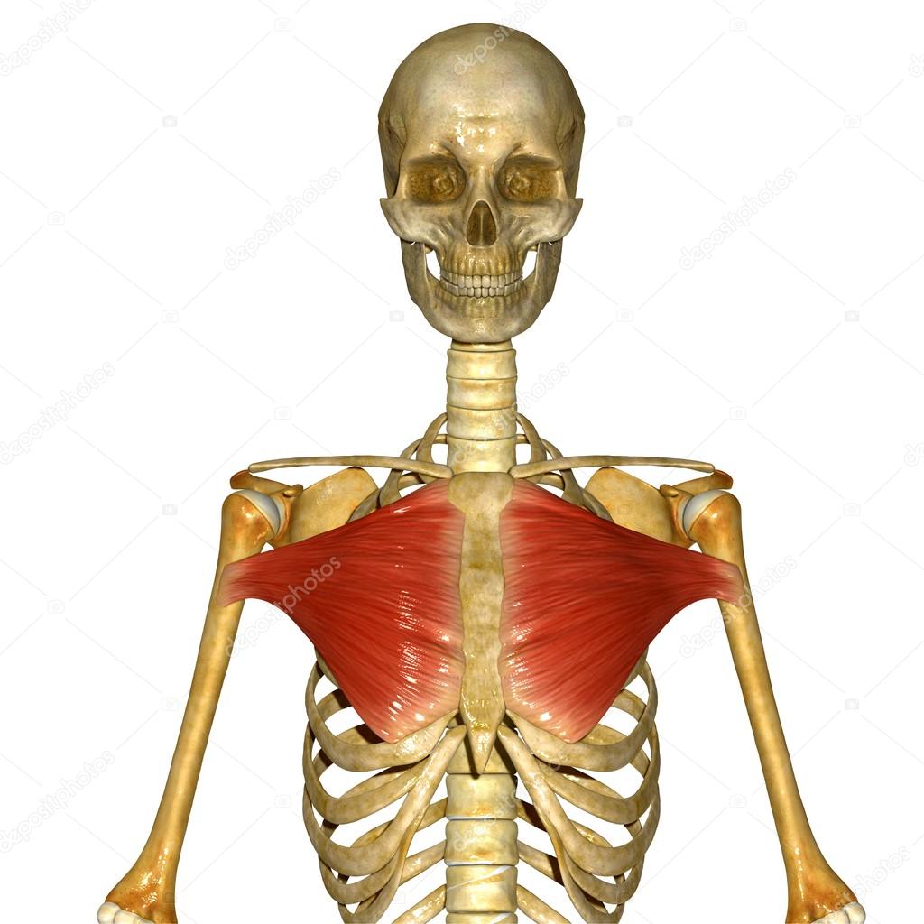
Computer artwork of a side view of some of the muscles (red) of the chest, showing their attachment to the bones of the ribcage and back. The pectoral region is located on the anterior chest wall. This page provides an overview of the chest muscle group. Except for the brain, the trunk houses all the vital organs of the human body. This muscle group is responsible for pushing movements and interacts synergistically with the anterior deltoid of the shoulder and triceps of the arm. This mri chest (thorax) axial cross sectional anatomy tool is absolutely free to use. Chest muscles anatomy the chest is made up primarily of two muscles: All about the chest muscles the chest anatomy includes the pectoralis major, pectoralis minor and the serratus anterior. Use the mouse scroll wheel to move the images up and down alternatively use the tiny arrows (>>) on both side of the image to move the images.>>) on both side of the image to move the images. It features dissection of the cat, numerous physiological experiments, and an emphasis on the study of anatomy through histology. See human chest anatomy stock video clips. Learn about each of these muscles, their locations, functional anatomy and exercises for them. Ventral trunk muscles (overview) the trunk (torso) is the central part of the body to which the head and the limbs are attached.